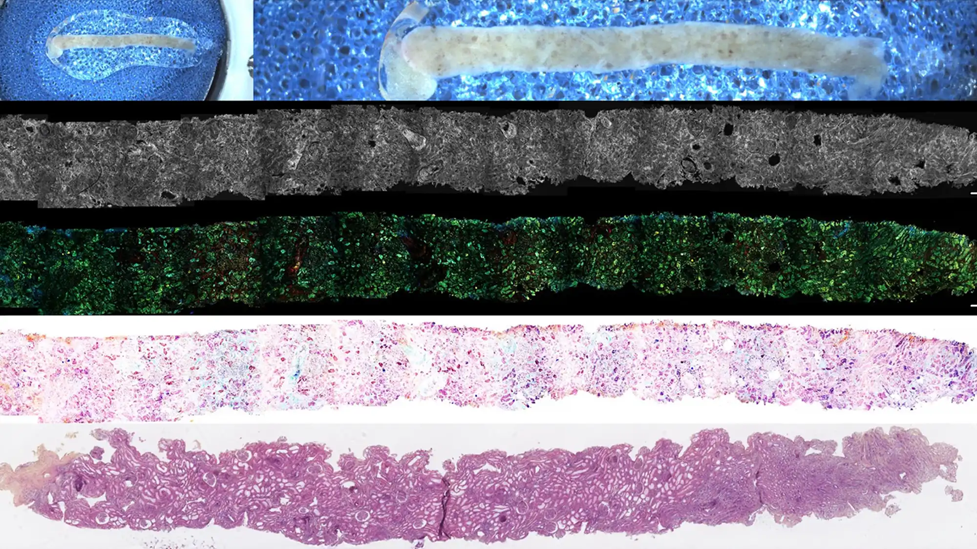
Rapid On-Site Kidney Biopsy Analysis With D-FF-OCT Imaging

Abstract
Background: Traditional histopathological analysis of kidney biopsies is critical for diagnosing and managing kidney disease but is often limited by time-consuming sample preparation. There is a need for faster, label-free alternatives to improve diagnostic efficiency and clinical decision-making.
Methods: This study evaluated dynamic full-field optical coherence tomography (D-FF-OCT) as a rapid, on-site imaging technique for kidney biopsy assessment. Thirty-one patients requiring either native or transplant kidney biopsies were included. Biopsies were analyzed using both conventional staining and D-FF-OCT imaging, with results compared for correlation and diagnostic accuracy.
Results: D-FF-OCT successfully identified major kidney structures and lesions such as interstitial fibrosis, tubular atrophy, and glomerular crescents without the need for extensive tissue processing. The degree of interstitial fibrosis and tubular atrophy assessed by D-FF-OCT correlated strongly with conventional histopathology (r = 0.61 and 0.60, P < 0.001). D-FF-OCT provided high-resolution, rapid images, enabling immediate evaluation of biopsy adequacy and preliminary pathology. The method demonstrated high concordance with standard stains and reduced the delay from biopsy to diagnostic insight.
Conclusions: D-FF-OCT is a promising, label-free technology that enables rapid, high-resolution on-site analysis of kidney biopsies. Its application may significantly accelerate diagnosis and treatment planning for both native and transplant kidney diseases, overcoming key limitations of traditional histopathological workflows .


