Use of the VanGogh System to Assess for Intra-Operative Tissue Adequacy: Initial Experience
Abstract
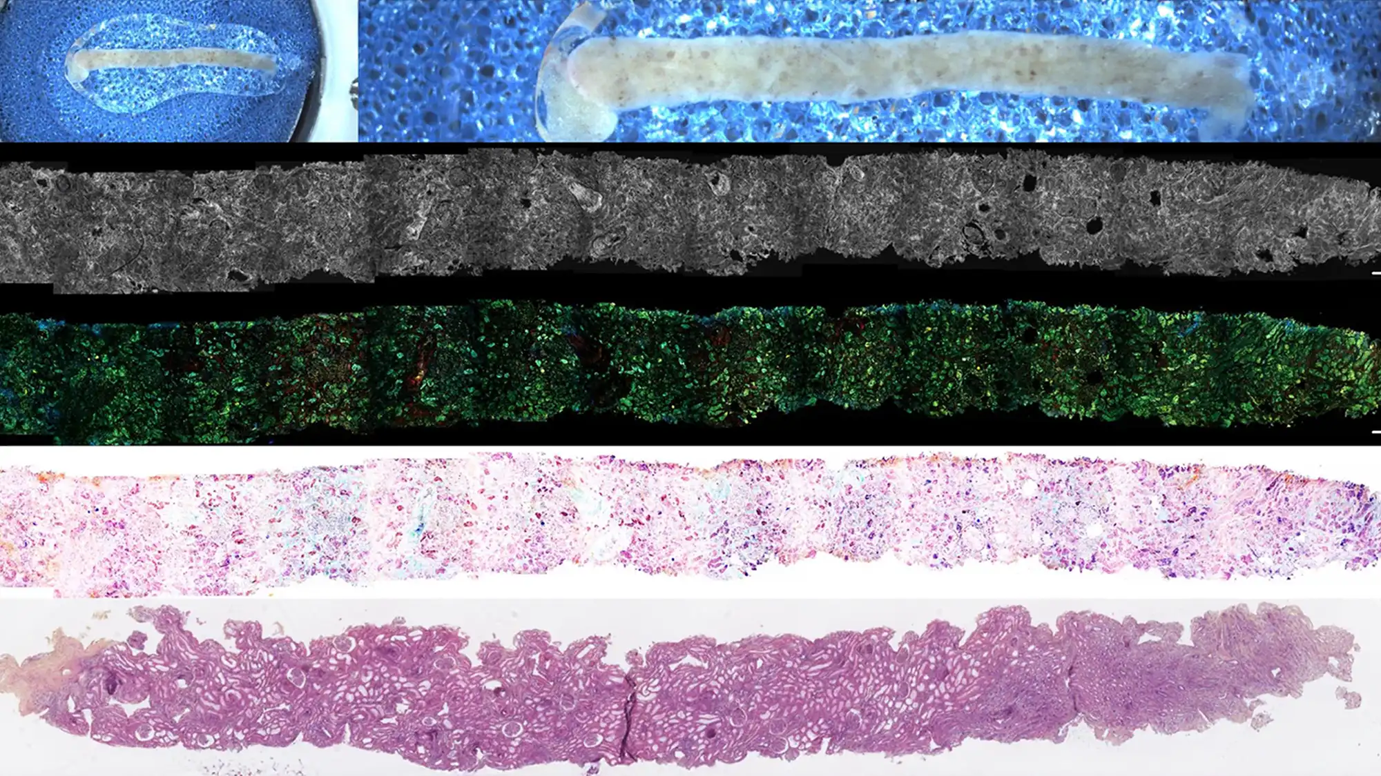
Background: Ensuring tissue adequacy during lung biopsy is critical for diagnosis and molecular testing, but conventional rapid on-site evaluation (ROSE) is limited by sample processing and partial analysis. Dynamic cell imaging (DCI), as used by the VanGogh system, enables real-time, label-free assessment of cellularity, morphology, and metabolic activity on unprocessed fresh tissue. This method also preserves specimens for molecular studies and is adaptable to machine learning for reproducible results.
Methods: In this initial experience, 51 peripheral lung biopsies were evaluated using the VanGogh system between March and September 2024. Biopsies were obtained via robotic bronchoscopy and scanned with the VanGogh platform to assess metabolic activity and lesional cell count. Final pathology results and adequacy for molecular profiling were analyzed.
Results: Of 51 biopsies, 24 were malignant and 27 were benign. Among 23 malignant cases assessed with the VanGogh system, 100% had adequate tissue for molecular profiling, a finding confirmed by subsequent formal pathology. Non-cancerous samples included infections and inflammatory lesions that resolved on follow-up.
Conclusions: Dynamic cell imaging with the VanGogh system provided rapid and accurate intra-operative assessment of lung biopsy adequacy. All malignant cases had sufficient material for downstream molecular testing, suggesting DCI may improve institutional efficiency and the reliability of lung cancer diagnostics .