Full-field OCT for High-Resolution Gut Biopsy Imaging
Abstract
Background: Advances in endoscopic imaging have significantly improved the diagnosis and management of gastrointestinal (GI) diseases. Full-field optical coherence tomography (FF-OCT) has emerged as a promising high-resolution imaging modality, enabling label-free, real-time visualization of GI mucosa and submucosa at near-histological detail.
Methods: This review synthesizes current research on the application of FF-OCT for GI tract imaging. The principles of FF-OCT technology are described, including its ability to generate en face and cross-sectional images with subcellular resolution. The review examines ex vivo and in vivo studies, technical developments, and the comparison of FF-OCT with traditional histology and other optical imaging modalities.
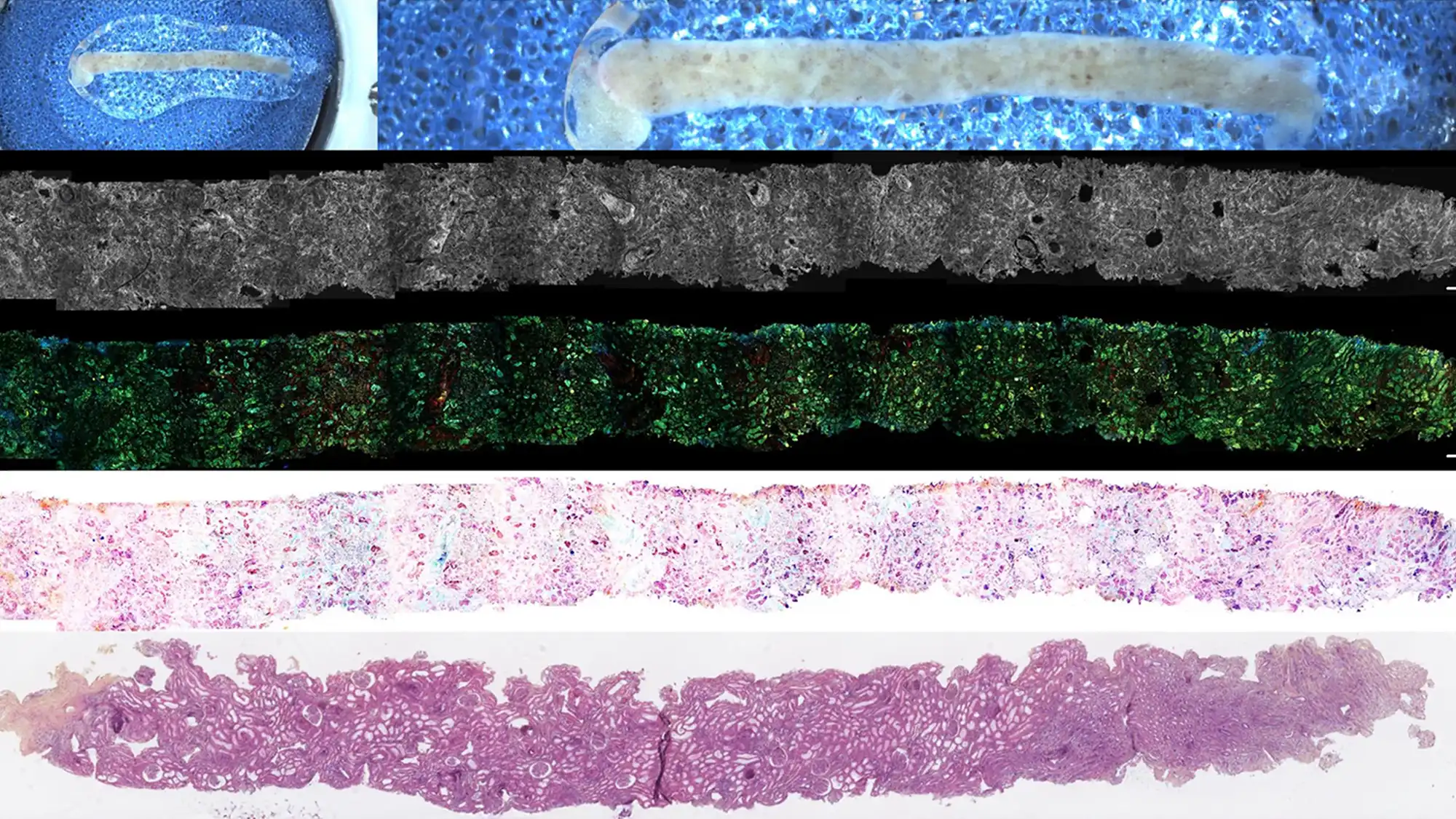
Results: FF-OCT provides high-resolution images of GI tissues, allowing for the identification of key histological features such as glandular structures, crypt architecture, and cellular morphology. Ex vivo studies have demonstrated strong correlation between FF-OCT images and standard histopathology. Early clinical investigations suggest the feasibility of in vivo FF-OCT imaging for real-time assessment of neoplasia, inflammation, and mucosal integrity. The technique is non-destructive, rapid, and does not require tissue staining.
Conclusions: FF-OCT represents a significant advancement in GI imaging, offering the potential for immediate, label-free microscopic evaluation of tissue during endoscopy or surgery. While ex vivo studies have established its diagnostic capabilities, ongoing technical refinements and clinical trials are needed to validate its routine use in vivo and to integrate FF-OCT into clinical workflows for GI disease diagnosis and management.