Use of the VanGogh System to Assess for Intra-Operative Tissue Adequacy: Initial Experience
Abstract
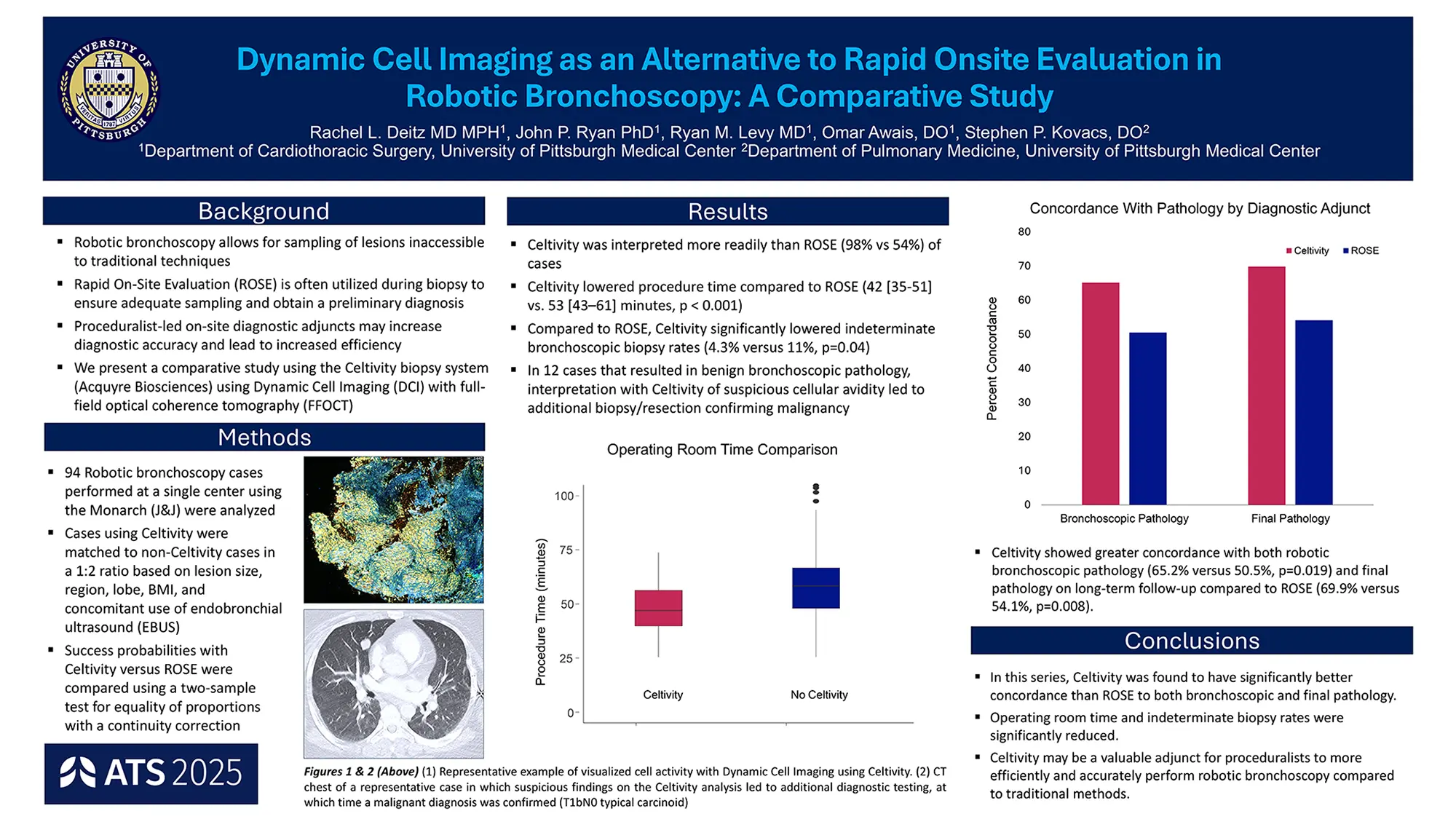
Background: As targeted cancer therapies demand more tissue for molecular analysis, traditional lung biopsies pose greater risks. Rapid on-site evaluation (ROSE) is limited, assessing only a small fraction of cells. Dynamic cell imaging (DCI) using the VanGogh system allows rapid, real-time evaluation of fresh tissue for cellularity, morphology, and metabolic activity while preserving the sample and supporting advanced analysis.
Methods: This study reviewed 51 peripheral lung biopsies obtained via robotic bronchoscopy and evaluated with the VanGogh system between March and September 2024. Samples were scanned, analyzed, and then assessed on final pathology.
Results: Of 51 biopsies, 24 were malignant, and 27 were benign. In all 23 malignant cases analyzed for molecular profiling, VanGogh achieved 100% adequacy. The technique enabled rapid, comprehensive assessment of lesional tissue.
Conclusions: Dynamic cell imaging with the VanGogh system rapidly and accurately determines lung biopsy tissue adequacy for molecular testing, potentially improving the safety, efficiency, and reliability of intra-operative cancer diagnostics .
Institution: University of Chicago Medicine