
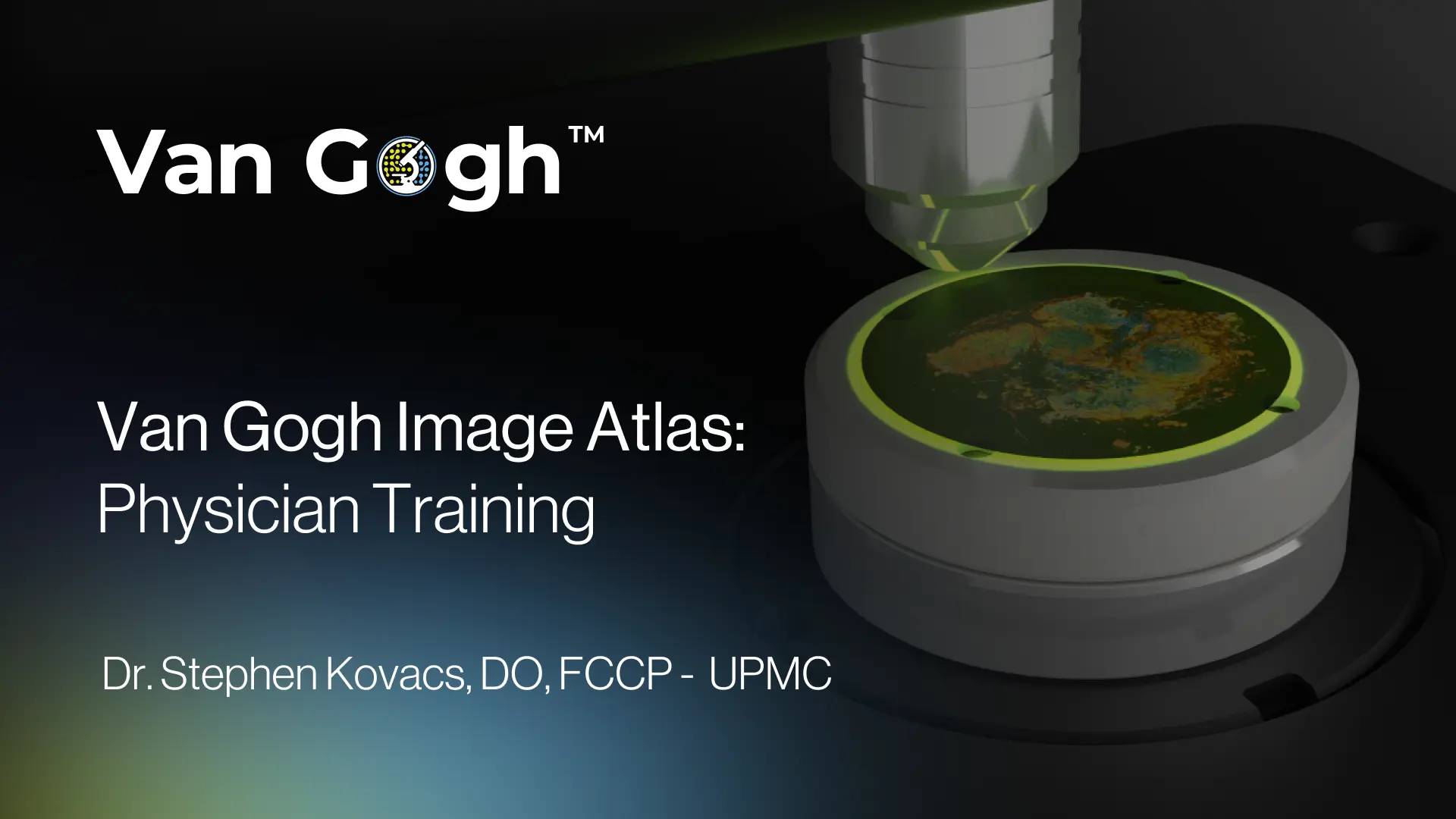
Van Gogh™ System: Educational Image Reference Guide

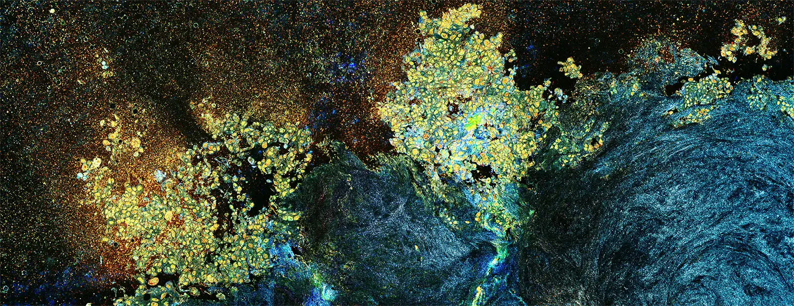
This educational reference guide demonstrates the visualization capabilities of the Van Gogh™ Microscopy System across a spectrum of tissue types. It provides side-by-side comparisons of Dynamic Cell Imaging™ (DCI), HistoView™, and Final Pathology results for samples collected via robotic bronchoscopy (both cryobiopsy and forceps).
Key Features:
- Visual Correlation: Illustrates the direct correlation between intraprocedural DCI images and final histopathology.
Tissue Types Covered:
- Benign: Normal lymphoid tissue (LN 7 FNA, LN 4R FNA).
- Inflammatory: Inflammation (RUL Forceps Biopsy).
- Infection: Pneumococcus infection (LUL Forceps Biopsy).
- Granuloma: Granuloma tissue (RLL Cryobiopsy, LN 7 FNA).
- Adenocarcinoma: Malignant tissue (RML Forceps Biopsy, LN 4L FNA).
- Educational Purpose: Designed to train physicians on interpreting DCI images to assess tissue adequacy and make informed intraoperative decisions.
Disclaimer: These images are for educational purposes to illustrate appearances observed with Van Gogh™ and are not intended for diagnostic determinations.
Our Mission
Empowering the proceduralist to improve patient outcomes through better diagnostic cancer sampling
Get in Touch
